Ultrasound
The information presented in this article has been gathered by Sofian Zeina and Abram Shihata and was presented in the Biomedical Engineering meeting on November 2017. The article is posted by Nikhil Kanamala.
Ultrasound waves are acoustic waves which are mechanical
vibrations inducing alternating refraction and compression of the medium
through which they pass. Ultrasound
waves have frequencies higher than the limit of the human hearing typically
between 1 – 20 MHz (generally humans can hear between 20 Hz and 20 KHz).
Acoustic waves contain the following properties:
·
Frequency
·
Wavelength
·
Amplitude
·
Propagation Velocity
Ultrasound waves interact with organs and tissues in the
human body with the following phenomenon (Figure 1):
·
Reflection
·
Scattering
·
Refraction
·
Attenuation

Figure 1: Ultrasound waves interaction with organs.
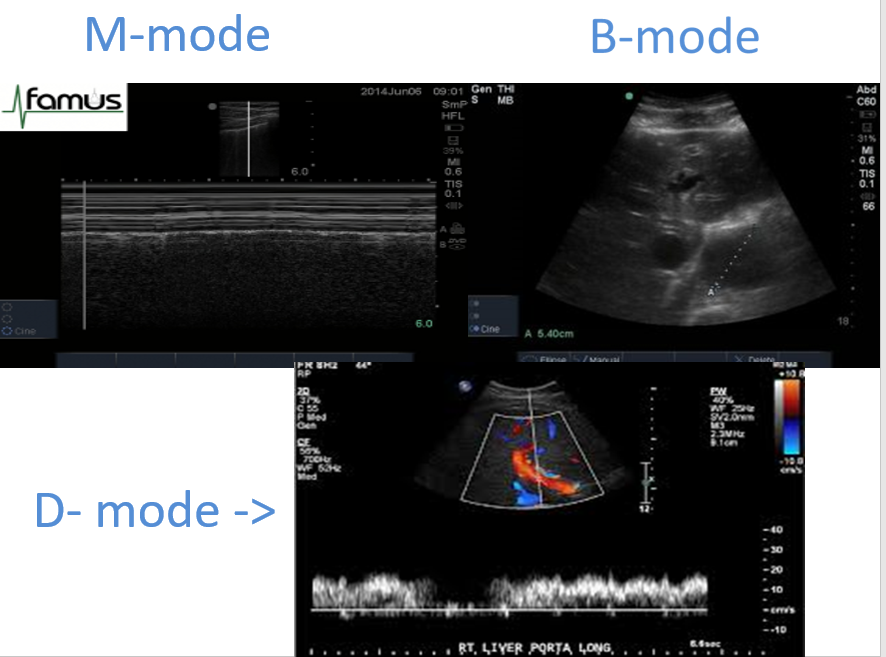
Ultrasound imaging contain the following modes:
·
Amplitude (A-mode) - For historical reference.
Amplitudes over time were used to show structural shifts. However, A-mode is
still in used for Eye ultrasound.
·
Motion (M-mode) - Reflects motion of the heart
structures over time. Used for accurate evaluation of rapid movements.
·
Brightness (B-mode) - Most essential modality.
Amplitude of reflected signal is converted into gray scale image.
·
Doppler (D-mode) - The Doppler effect describes
a change in frequency of sound waves depending on the direction in which
they’re travelling (and being recorded).
Doppler (D-mode) contains the following subsections:
·
Doppler Duplex - Based on the simultaneous
B-Mode and Doppler imaging
·
Continuous-Wave Doppler (CWD) – Defines blood
flow direction, very useful in high velocity signals recording.
·
Pulsed–Wave Doppler (PWD)- Provides both blood
flow direction and precise determination of Doppler signal source.
·
Color Doppler – Based on the PWD. Velocities are
displayed using color scale. Velocities toward the transducer are red, and
velocities away from the transducer are blue.
Ultrasound Transducers construction vary based on the following:
·
Piezoelectric crystal arrangements
·
Aperture (footprint)
·
Operating frequency (related to the penetration
depth)
The most popular types of Ultrasound Transducers are:
·
Sector transducer- mainly echocardiography, gynecological,
upper body ultrasound
·
Linear transducer – mainly obstetric, breast,
thyroid and vascular ultrasound
·
Convex transducer – mainly in all ultrasound
types except echocardiography
Implementing multiple point of care
ultrasound imaging devices (S-series, Edge II, M-Turbo and Logiq E) has the following advantages:
·
Portability
·
Affordability
·
Time Saving
·
Simplicity
Fusion Ultrasound has combined previously acquired CT, PET
or MRI images with real time ultrasound.
However, it does require compatible hardware and software. The advantages are:
•
More data helps users localize areas of interest.
•
Greater accuracy (either in needle navigation or
in diagnostics).
•
Procedure time is reduced
•
Portable

If you are searching for the best IVF Center in Surat, Devaki IVF Centre is a premier choice for fertility treatment in Surat. Our IVF specialist use sonography to check internal health for a successful pregnancy. Sonography is a type of medical test that uses sound waves to take pictures inside your body.
ReplyDelete